Biology students blend science and creativity through anatomy-inspired art
Share

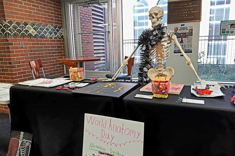
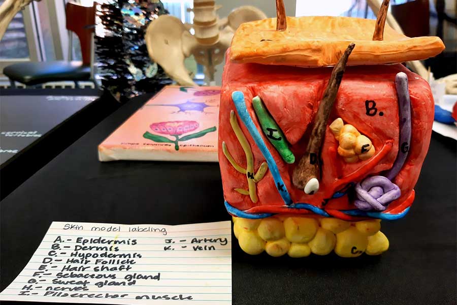
Students in Marshall University’s Human Anatomy Laboratory explored the intersection of science and art through a collection of anatomy-inspired projects that showcased their creativity and understanding of the human body.

The projects included detailed anatomical illustrations, creative 3D models, and other original works inspired by concepts studied in the laboratory. By translating scientific knowledge into visual and artistic forms, students demonstrated both their mastery of course material and their creative talents.

The collection highlights the many ways students can engage with anatomy beyond the traditional classroom, using art to communicate complex biological concepts and celebrate the study of human anatomy.
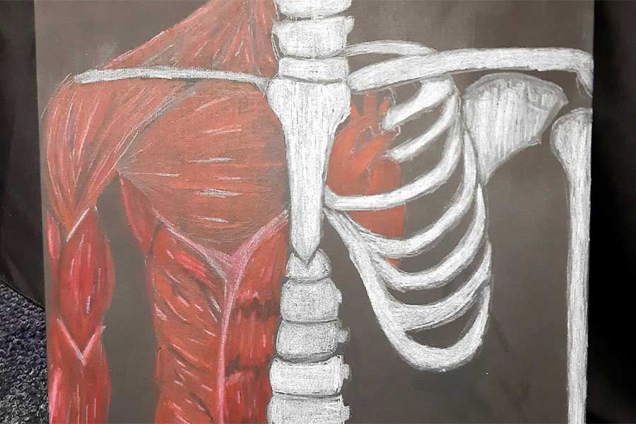
Close-up of a student artwork depicting human anatomy, with muscles illustrated on one side of the torso and skeletal structures, including ribs and part of the spine, shown on the other side. A red heart is visible within the rib cage against a dark background.

Close-up of a student-created 3D skin model showing layers of the skin, hair follicles, nerves, blood vessels, and glands. A handwritten key identifies anatomical structures displayed in the colorful cross-sectional model.

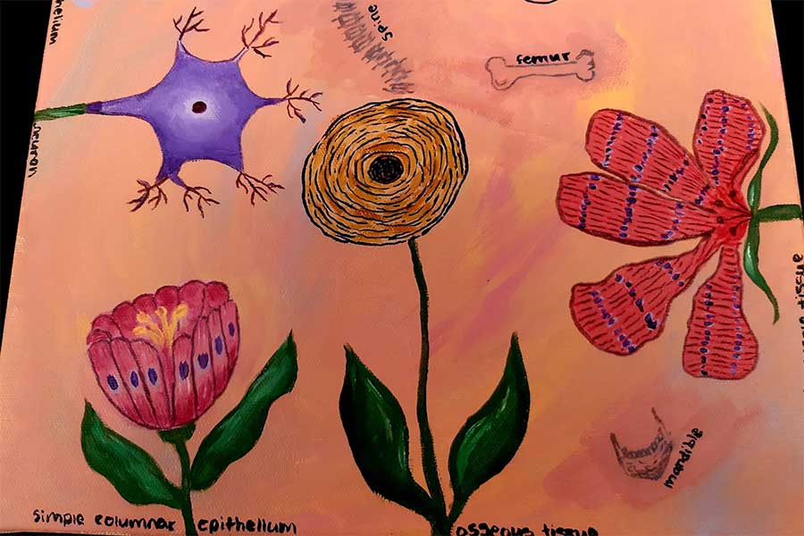
Student artwork combines anatomy and biology concepts by depicting cells, tissues, and anatomical structures as flowers. Visible illustrations include a neuron, compact bone tissue, simple columnar epithelium, skeletal muscle tissue, and a labeled femur against a pink background.

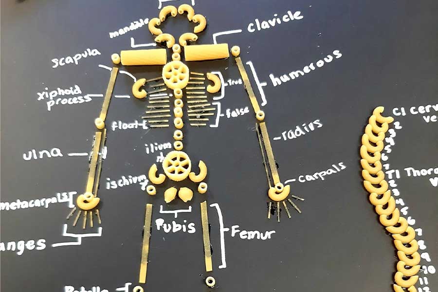
Student anatomy project uses pasta and other materials to create a labeled model of the human skeleton and vertebral column. Anatomical terms including scapula, clavicle, humerus, radius, ulna, pubis, femur, and cervical vertebrae are written around the handmade display on a black background.